Surgical Precision
Today we scrubbed into the Coastal Resource lab at Walailak University and familiarized ourselves with different marine life anatomy. The first aquatic friend we cut open was a Japanese Threadfin Bream. To begin our dissection we studied the exterior of the fish in order to know where to make our first cuts and where to find the interior masses. The first bream was a male indicated by the large gonads (fig. 3), in addition we found a Silver side in his stomach (fig.4) although it was mostly digested and lost most of it’s distinguishing features.

- (fig.1) Japanese Threadfin Bream Nemipterus japonicus

- (fig.2)

- (fig.3) Fish Gonads

- (fig.4) Silver side fish nearly fully digested by the Japanese Threadfin Bream found in it’s stomach
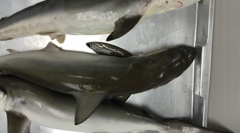
After the fish we moved on to something larger and began to dissect a shark. This particular shark is a Spot Tail and was a male, see fig. 7. He was very meaty with two large livers lining his torso shown in fig. 8 and his intestines were found outside of his anus before we had even begun. This shark had tough skin almost too tough for our tools.

- (fig.5) Spot Tail Shark Carcharhinus Sorrah
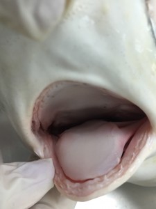
- (fig.6) This species has no spiracle, which functions like the marine version of nose, so it just takes water from its mouth

- (fig.7) These are the claspers, which identifies this shark as a male.
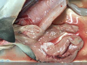
- (fig.8) This shark has two livers.

- (fig.9)

- (fig.10) This sharks heart has two valves.
We finished our dissection experience by examining a Green Mussel, they were the only specimen studied during the lab that wasn’t caught fresh that day. The hardest part was finding a way to crack open the right and left valves to open the mussel. From there we could then find the major muscle and the foot.

- Green Mussel Perna viridis

It was very interesting to see how these animals bodies are so different from one another as well as so different from humans in order to survive in their habitats and preform their ecosystem functions.